
Figure 1.
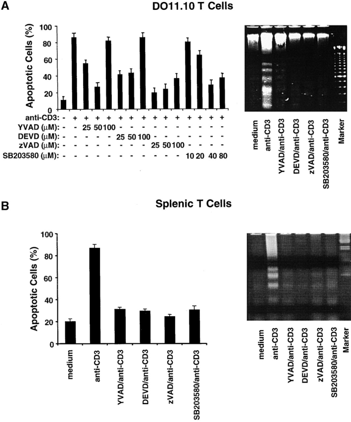
p38 MAPK is required for T cell AICD. (A) DO11.10 T hybridoma cells were pretreated with different concentrations of SB203580 or caspase inhibitors YVAD, DEVD, and zVAD and then stimulated with plate-bound anti-CD3 mAb (10 μg/ml) for 16 h. The number of dead cells was quantitated by PI staining, and the results are expressed as percent apoptotic cells (left panel). Data are shown as mean values ± SEM and are from one of two independent experiments. DO11.10 T hybridoma cells were pretreated and then stimulated as in A. Chromosomal DNA was extracted and analyzed on a 2% agarose gel to detect DNA fragmentation (right panel). (B) Purified splenic T cells from B6 mice were activated with plate-bound anti-CD3 and anti-CD28 mAbs for 48 h. Activated T cells were then restimulated in wells coated with anti-CD3 for 16 h in the presence of either the caspase inhibitors YVAD, DEVD, or zVAD or the p38 MAPK inhibitor SB203580. rIL-2 (20 U/ml) was added to unstimulated and restimulated cells. The percent apoptotic cells was determined by PI staining (left panel). Data are shown as mean values ± SEM and are from one of three independent and reproducible experiments. DNA fragmentation (right panel) was detected as in A. Data shown are from one of three independent and reproducible experiments.