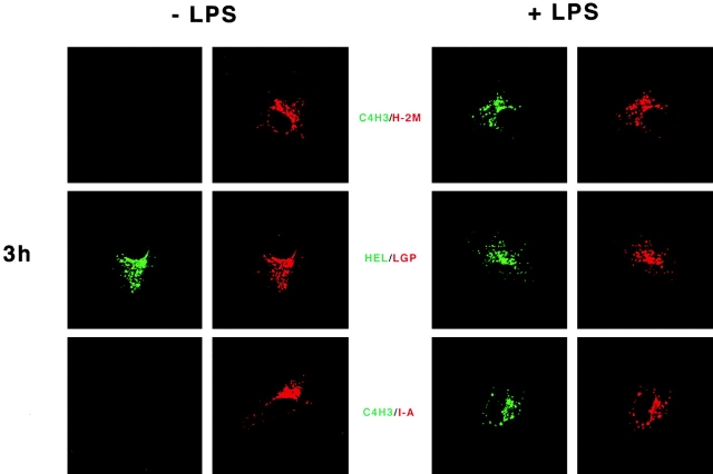
Figure 3.
Localization of HEL protein and MHC class II–peptide complexes in DCs. The procedure was the same as described in the legend to Fig. 2, but here the formation of MHC class II–peptide was monitored at 3 and 24 h by immunofluorescence confocal microscopy. On the left are CBA DCs cultured in HEL only, and on the right are cells cultured with HEL + LPS. Representative cells are shown following double labeling in green for the HEL antigen, either HEL protein with 1B12 antibody or I-Ak/HEL MHC-peptide complexes with C4H3. Red stain identifies the lysosomal membrane glycoprotein lysosomal-associated membrane protein 2. Identical red staining is seen with H-2M or MHC class II. The results are representative of >10 experiments, and of experiments with both HEL protein and preprocessed HEL peptide. The maturation-induced C4H3 signal at 24 h is shown for a well-spread cell, but in optical sections through thicker cells, C4H3 stain is almost entirely along the cell perimeter.


