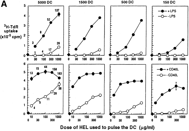
Figure 5.
Antigen presenting activity of DCs in vitro after culture with graded doses of HEL, minus or plus LPS or CD40L as a maturation stimulus. (A) Day 6 bone marrow DCs from CBA mice (top) or C3H/HeJ mice (bottom) were cultured for 20 h with graded doses of HEL (x-axis) that had been endotoxin depleted with Kuttsuclean™. One group of cultures was matured by simultaneous addition of LPS or CD40L (closed symbols), and the other was unstimulated (open symbols). After 20 h, the cells were harvested, washed, and fixed in paraformaldehyde to block further processing and maturation. MHC–peptide complexes were quantified in terms of C4H3 staining, and are shown as mean fluorescence indices on the left. The fixed DCs were added in graded doses (top) to 250,000 CD4+ T cells from 3A9 TCR transgenic mice (specific for the same complex of I-Ak + HEL peptide as C4H3 antibody). 3H-TdR uptake was measured at 30–42 h. One of three similar experiments. (B) The display of TCR ligands by fixed DCs was monitored at 5 h on T cells (gated away from DCs by light scattering) by the criteria of TCR downregulation (anti-Vβ8, y-axis) and CD69 upregulation (x-axis). The DC/T cell ratio was 1:3 in the data that are shown, and the percentage of CD69+ cells is indicated in each dot plot (upper right).


