
Figure 5.

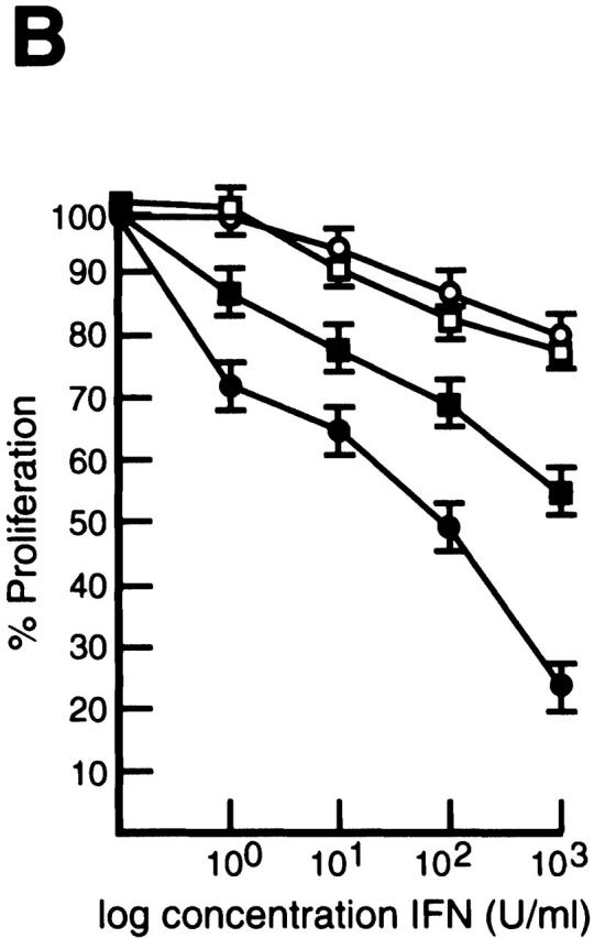


Constitutive IRF-4 expression promotes c-rel −/− B cell proliferation in the presence of IFNs. (A) IRF-4 is not expressed in c-rel −/− B cell lines. 5 μg samples of total RNA isolated from the immortalized B cell lines W404.1 (c-rel +/+; lane 1) and B1.1 (c-rel −/−; lane 2) were analyzed by Northern blot hybridization. Filters were sequentially hybridized with murine IRF-4 and rat GAPDH cDNA probes and exposed for autoradiography for 24 h. The results obtained with these two cell lines were representative of other independent c-rel +/+ and c-rel −/− cell lines. (B) c-rel −/− B cell lines are more sensitive to the antiproliferative activity of IFNs. The W404.1 (c-rel +/+; open symbols) and B1.1 (c-rel −/−; filled symbols) B cell lines were either untreated or stimulated for 72 h with 1, 10, 100, or 1,000 U/ml of IFN-α (circles) or IFN-γ (squares). Cellular proliferation was measured at 24-h intervals over 72 h by [3H]thymidine incorporation. Proliferation of all cell lines in the presence of a particular concentration of IFN at the 72-h time point is represented in this figure as a percentage of [3H]thymidine incorporated by normal or c-rel −/− cells, respectively, in the absence of IFN. All results represent the mean ± SD from four experiments. (C) Expression of exogeneous IRF-4. Whole cell extracts from W404.1 (c-rel +/+; lanes 1 and 2) and B1.1 (c-rel −/−; lanes 3 and 4) cells that had been infected with a control retrovirus (lanes 1 and 3) or a retrovirus expressing NH2-terminally FLAG-tagged IRF-4 (lanes 2 and 4) were resolved by SDS-PAGE and subjected to Western blot analysis using mAbs directed to the FLAG epitope. (D) Enforced IRF-4 expression overcomes the heightened anti-proliferative action of IFNs that result from the loss of Rel. The cell lines W404.1 (circles) and B1.1 (triangles) that were infected with a control retrovirus (open symbols) or a virus expressing FLAG-tagged IRF-4 (filled symbols) were either untreated or stimulated for 72 h with 1, 10, 100, or 1,000 U/ml of IFN-α or IFN-γ. The data shown represents the 72-h time point and are expressed as a percentage of [3H]thymidine incorporated by normal or c-rel −/− cells, respectively, in the absence of IFN. All results represent the mean ± SD from four experiments.