Figure 6.
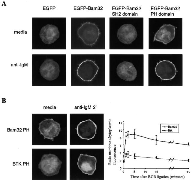
Bam32 is recruited to the plasma membrane after BCR ligation through its PH domain. BJAB B cells were electroporated with constructs encoding EGFP fused to Bam32 or individual domains of Bam32. 18–20 h after transfection, cells were harvested and replated in medium containing 1.25% FCS. The next day, cells were harvested, stimulated with anti-IgM F(ab′)2 fragments for 5 min, fixed, and mounted on slides. EGFP fluorescence was visualized using a scanning laser confocal microscope. (A) Bam32 associates with the plasma membrane through its PH domain. (B) Membrane recruitment kinetics of the Bam32 PH domain versus the BTK PH domain. Digital images of EGFP fluorescence were used to determine the ratio of membrane to cytoplasmic fluorescence intensity, as described in Materials and Methods. The graph indicates the average and standard error for six to nine cells per point, pooled from two anti-IgM stimulation experiments. Images representative of those used to generate the quantitative data are shown.

