Figure 3.
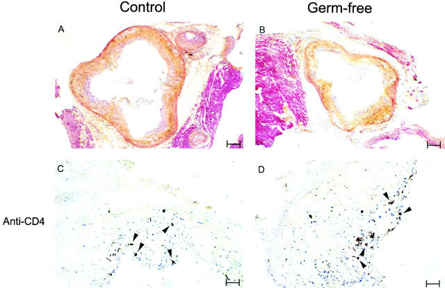
Histology of aortic root lesions in germ-free and control apo E−/− mice. (A and B) Cryosections of the aortic root area in mice at 22 wk of age were stained with hematoxylin-phyloxine-saffron. Lesions in both types of animal have fibrous components (indicated by yellow staining) and large areas that are rich in foam cells (indicated by light purple staining). White rice grain–shaped spaces indicative of the extracellular deposition of cholesterol are also visible in lesions from control and germ-free mice. The tissue staining red is surrounding muscle. Bar = 156 μm. (C and D) These sections are stained brown for the T lymphocyte marker CD4. Arrowheads indicate the location of T lymphocytes, present in both control and germ-free animals. Bar = 31 μm. Hearts from five mice of each type for both males and females were examined histologically, and the sections shown are representative of the results from all of the mice. The same morphology and cellular composition was observed in both female and male mice. Histology of hearts from two mice of each type at 32 wk of age similarly revealed no differences between germ-free apo E−/− and control apo E−/− mice (not shown). Additionally, staining for the macrophage marker CD11b revealed comparably stained areas in control and germ-free animals (not shown).

