
Figure 2.
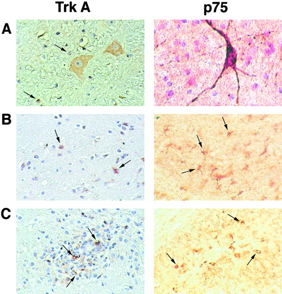
Characterization of NGF receptors in marmoset brain by immunohistochemistry. Staining for TrkA is shown at the left (5-μm paraffin-embedded section), and for p75NGFR at the right (30-μm-thick section stained in flotation). (A) Both NGF receptors are present in cholinergic neurons of the basal forebrain. Arrows show satellite oligodendrocytes stained for TrkA (left). (B) Arrows indicate some of the TrkA- and p75NGFR-positive glial cells (astrocytes or microglia) in normal white matter. (C) In CNS inflammatory infiltrates, strong staining for both NGF receptors is also apparent on many mononuclear cells and macrophages (arrows). Original magnifications: ×400.