
Figure 2.
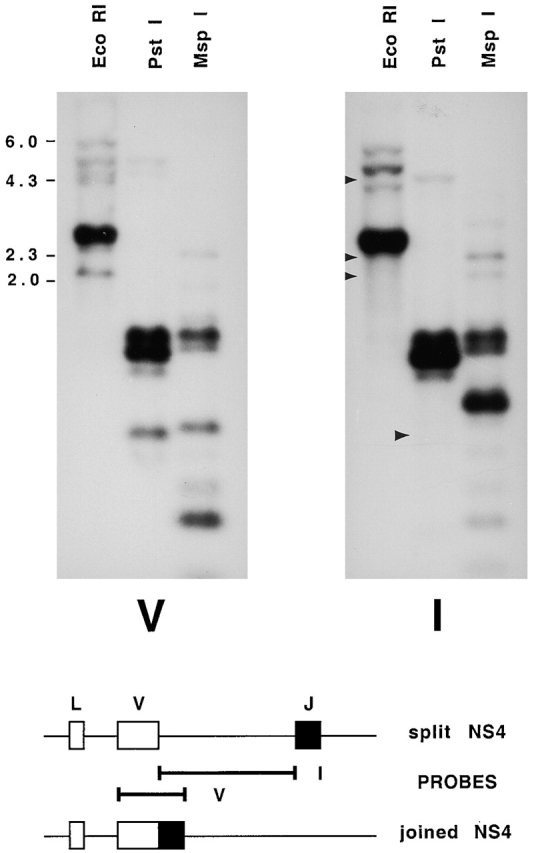
Genomic Southern blot hybridized with probes to the coding and intervening regions of NS4. Top, erythrocyte DNA from shark Y was digested with EcoRI, electrophoresed on a 1.2% agarose gel, and transferred onto a nylon filter. The filters were hybridized with radiolabeled DNA sequences from the V region of NS4 (V) and the intervening sequence between V and J (I). Arrows in the EcoRI lane point to the location of V-hybridizing bands at 2.1, 2.4, and 4.6 kb that were not detected by the I probe. These were cloned and contained germline-rearranged NS4 genes. Arrow in the PstI lane points to a fragment of 727–744 bp containing joined genes R4, R7, R18, R6, and R19, predicted from DNA sequence in Fig. 3 (dots over positions 79 and ∼816); accordingly, this fragment does not hybridize to I probe. The slower migrating bands in the same lanes at ∼1,150, 1,250, and 1,400 bp correspond to sizes expected with the presence of an intervening sequence of ∼500 bp; these bands accordingly hybridized with both V and I probes. Molecular size markers from λ phage DNA digested with HindIII are shown. The PstI fragment sizes were calculated from another gel; not shown. Bottom, location of V and I probes from cloned split (S5) and joined (R4) genes.