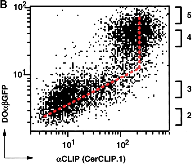
Figure 1.
DOαβGFP affects peptide presentation by HLA-DR3. (A) FACS® analysis of 5,000 Mel JuSo cells transfected either with DOαβ2 GFP (GFP+ population) or vector only (GFP− population) showing staining with secondary antibody only (−), or staining that is specific for class II molecules (L243), CLIP-bound HLA-DR3 (CerCLIP.1), or HLA-DR3 complexed to stably bound peptides (16.23). (B) FACS® analysis of 5,000 Mel JuSo cells expressing various levels of DOαβGFP, showing specific staining of HLA-DR3–CLIP complexes by CerCLIP.1 antibody. The vertical axis represents GFP and the horizontal axis PE-fluorescence derived from the secondary antibody, each in arbitrary units on a logarithmic scale. Values next to the right axis indicate which DOαβGFP populations were FACS® sorted for analysis of relative DO/DM expression levels.


