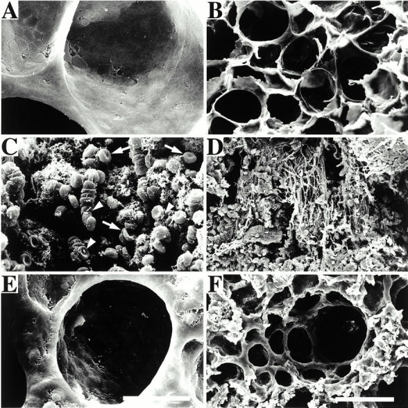
Figure 9.

Scanning electron microscopy of rat lungs. The figure shows micrographs of glutaraldehyde-fixed lungs from an uninfected rat (A and B), a rat infected with SR11B bacteria (C and D), and a rat infected with SR11B bacteria and treated with H-D-Pro-Phe-Arg-CMK (E and F). Arrows point to erythrocytes and arrowheads to bacteria adhering to erythrocytes. Bars, 10 μm (A, C, and E) and 100 μm (B, D, and F).
