Figure 4.
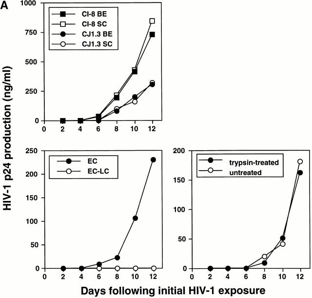
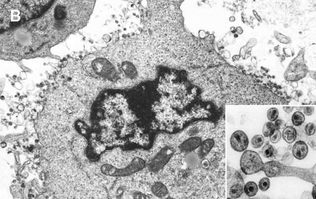
HIV-1–infected LCs are responsible for transmitting high levels of infection to cocultured CD4+ T cells. (A, top left) Tissue explants were exposed to two HIV-1 primary isolates, HIV-1CJI.3 (R5) or HIV-1CI-8 (R5X4), on the basal epithelial (BE) side or the stratum corneum (SC) side, washed, and then floated in media containing allogeneic CD4+ T cells. (B) electron micrograph taken from an LC–T cell coculture 7 d after HIV-1CI-8 exposure of explants showing numerous HIV-1 virions surrounding and budding from a CD4+ T cell (×64,000; inset ×440,000). (A, bottom left) Epidermal cell suspensions were made immediately after HIV-1 exposure of epithelial tissue explants to HIV-1CJI.3, and half of the suspensions were then depleted of LCs by immunomagnetic bead separation. Nondepleted epidermal cells (EC) and LC-depleted epidermal cells (EC-LC) were then cocultured with allogeneic CD4+ T cells. (A, bottom right) Emigrated LCs were collected from media 2 d after HIV-1CJI.3 exposure of epithelial tissue explants, and half of the cells were exposed to trypsin treatment and the other half left untreated. Trypsin-treated LCs and untreated LCs were then cocultured with allogeneic CD4+ T cells. HIV-1 infection levels were assessed by measuring HIV-1 p24 protein content by ELISA in coculture supernatants collected every other day (A, all panels). Data shown are representative of at least three separate experiments that showed similar results.


