Figure 5.
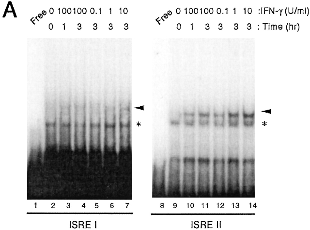
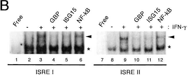
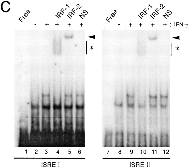
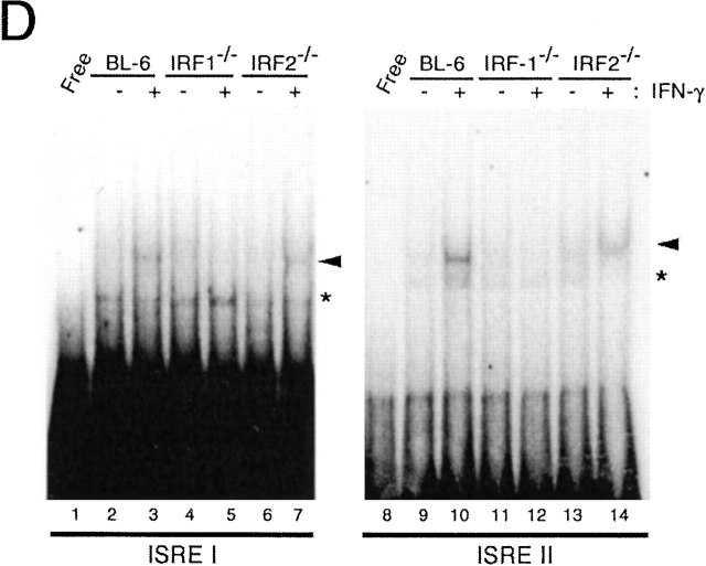
IRF-1 and IRF-2 bind to both Cox-2 ISREs. (A) EMSAs performed using probes containing the ISRE I and ISRE II from the murine Cox-2 promoter and nuclear extracts from C57BL/6 macrophages incubated with IFN-γ for different periods of time or doses, as indicated. “Free” lanes represent the mobility of the probe without any extract added. Black arrowheads point to the IFN-γ–inducible complex. Asterisks indicate the constitutive complex. (B) DNA binding specificity experiments on ISRE I and ISRE II using for competition 50-fold excess of cold ISREs (GBP- ISRE; lanes 4 and 10, and ISG15-ISRE; lanes 5 and 11) or a nonspecific competitor (NF-κB binding site; lanes 8 and 12). (C) Supershift experiments on ISRE I and II using IRF-1– or IRF-2–specific antibodies as indicated. NS, nonspecific antiserum. (D) EMSA using control C57BL/6+/+ or IRF-1−/− and IRF-2−/− macrophages.


