
Figure 3.
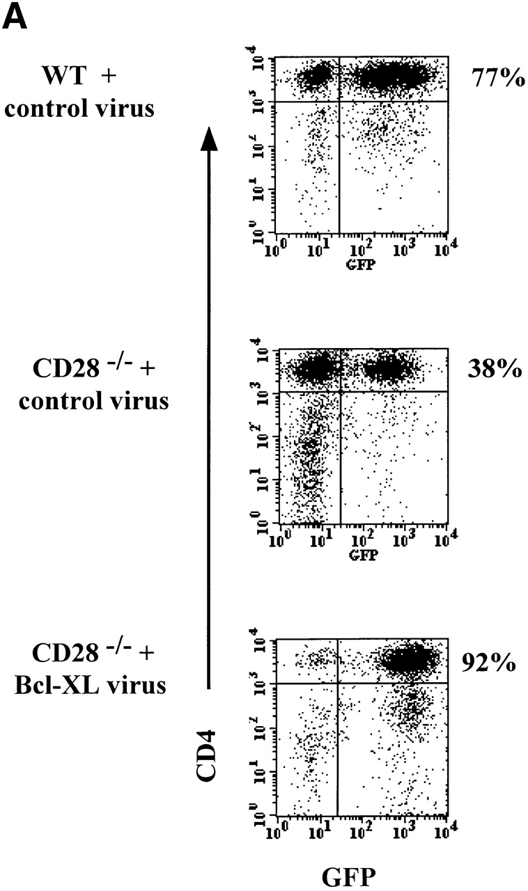

Expression of Bcl-XL in CD28−/− T cells does not restore normal proliferation. DO.11 WT or DO.11 CD28−/− T cells activated in vitro were infected with control or Bcl-XL virus on day 1 (as described in the legend to Fig. 1) and harvested 3 d later. Using a Cytomation MoFlo high speed sorter, the CD4+KJ1-26+ cells expressing high levels of GFP were collected. The cells were then plated in 24-well plates at 2.5 × 10⁵ T cells/well with 2.5 × 10⁶ APCs and 1 μg/ml peptide or in 96-well plates at 2.5 × 10⁴ T cells/well with 2.5 × 10⁵ APCs and a range of OVA peptide concentrations. (A) After 4 d of culture in 24-well plates with APCs and OVA peptide, the cells were collected and stained with antibodies to CD4. The cells were then analyzed by flow cytometry for GFP expression in the CD4+ population. (B) The microcultures of DO.11 WT or DO.11 CD28−/− GFPhigh T cells fractions stimulated with APCs and OVA peptide were pulsed with [3H]thymidine for 6 h on days 1 and 3, and the incorporated radioactivity was measured.