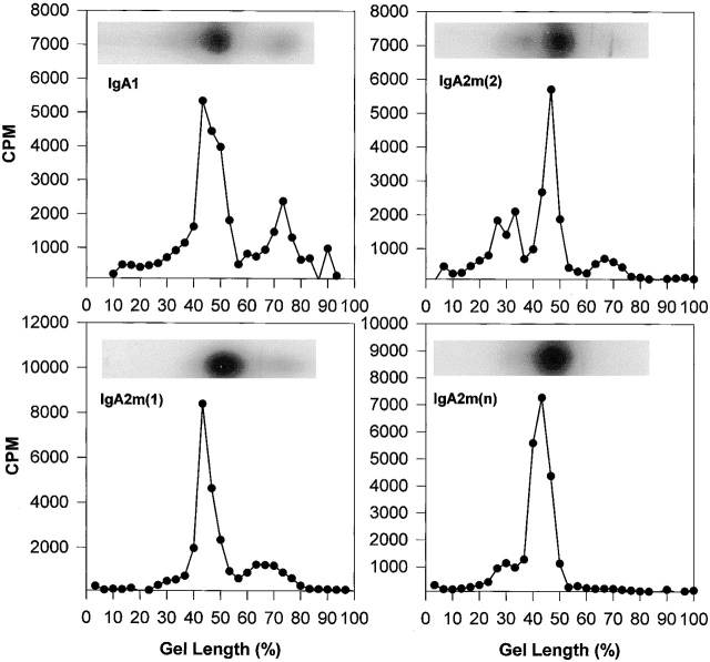
Figure 1.
Analysis of IgA1, IgA2m(1), IgA2m(2), and IgA2(n) by native gradient PAGE. To determine the monomer, dimer, and polymer composition of IgA proteins, proteins were analyzed on 2–16% native polyacrylamide slab gels in the absence of any denaturant. An autoradiogram of each IgA preparation shows the distribution of radioactivity in the gel channel. For quantitative analysis, the gel channels were sliced and counted as described in Materials and Methods. Monomeric IgA migrates furthest and localizes toward the bottom of the gel between 60 and 80% of the gel length. IgA dimer migrates and localizes between 35 and 50% of the gel length.

