
Figure 4.
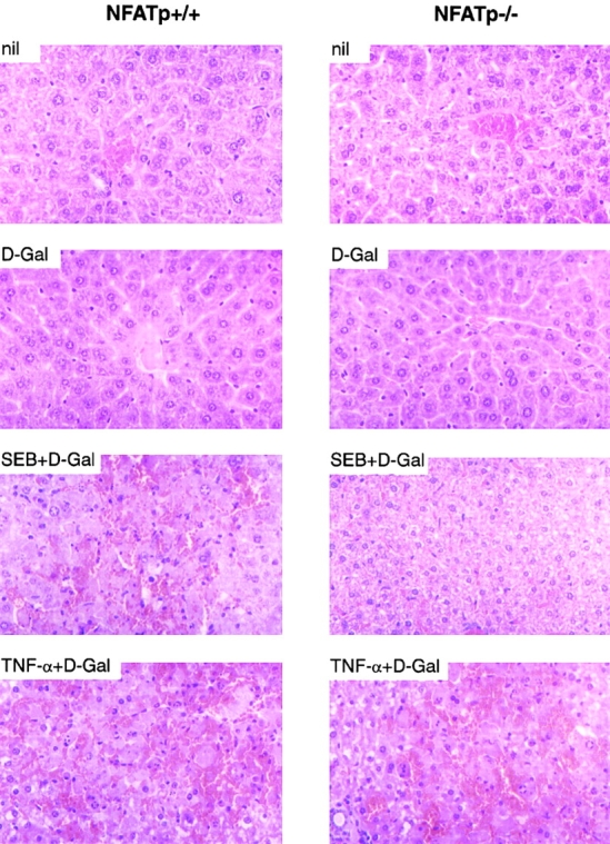
Histological analysis of liver sections after superantigen or TNF-α challenge. Liver was removed from NFATp+/+ and NFATp−/− mice 8 h after intraperitoneal injection of 0.9% NaCl (nil), D-Gal (20 mg), SEB (5 μg) plus D-Gal, or recombinant TNF-α (1 μg) plus D-Gal as indicated. Tissues were directly transferred into 10% formalin solution. Sections were stained with hematoxylin and eosin. Original magnification: approximately ×100.