Figure 7.
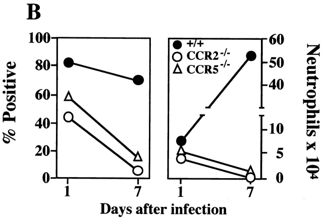
Marked neutrophilic inflammation in mice deficient in CCR2. (A) The histopathological sections (hematoxylin and eosin; original magnification: ×400) are from mice infected for 5 wk with L. major. Abundant organisms admixed with areas of necrosis, karryohectic cellular fragments, and polymorphonuclear leukocytes including eosinophils were found in CCR2−/− mice. In contrast, the ears from +/+, CCR5−/−, and MIP-1α−/− mice had fibroblasts with scattered cellular infiltrates that consisted of mononuclear cells, epitheloid histiocytes, lymphocytes, and plasmacytoid cells. Necrosis was absent in these three groups of mice. (B) Marked recruitment of neutrophils to the ears of CCR2-null mice after intradermal infection with L. major. 1 and 7 d after L. major infection, the cells that emigrated from ear skin explants were stained with FITC-labeled anti-I-Ab mAb and PE-labeled anti–Ly-6G mAb. Percentage (left) and total number (right) of neutrophils (Ly-6G brightI-Ab−) among all cells that emigrated from skin explants of +/+ (○), CCR2−/− (•), and CCR5−/− (▵) mice are shown. Results from one of three representative experiments are shown.


