
Figure 3.
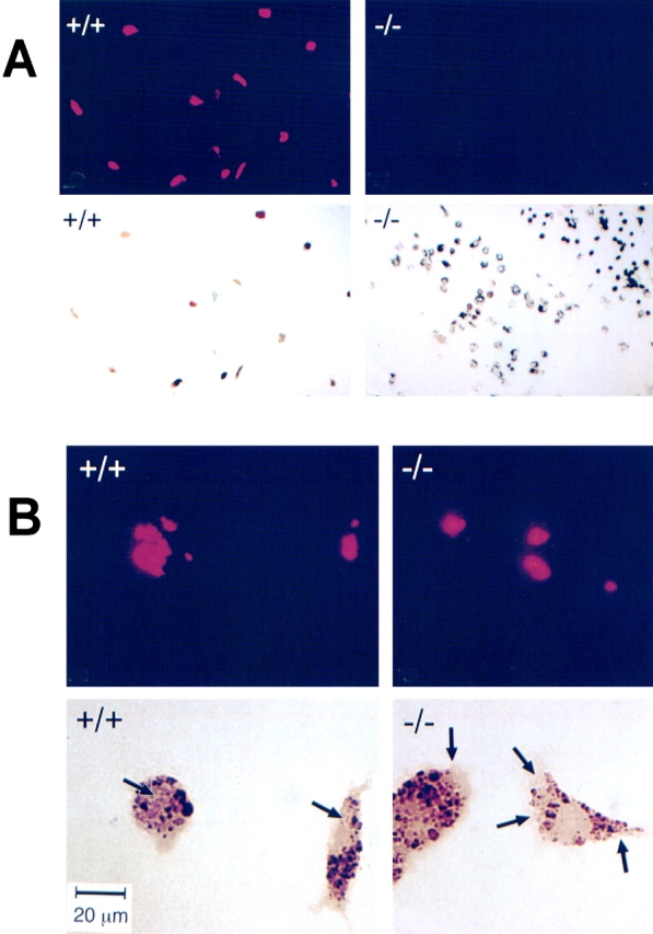
Association of M. avium with CD43+/+ and CD43−/− Mφ examined by fluorescence microscopy. M. avium were incubated with Mφ at a ratio of 20:1 for 4 h. After washing and fixing, mycobacteria associated with the cell monolayers were stained with auramine-rhodamine (TB staining reagents), and the preparations were examined by microscopy. (A) Shown are fluorescent micrographs (top) and phase–contrast images of the same frames (bottom) of CD43+/+ (left) and CD43−/− (right) Mφ. Note that almost all of the CD43+/+ Mφ have multiple associated mycobacteria; in contrast, negligible numbers of mycobacteria are associated with CD43−/− Mφ. (B) Higher magnification images. Description as in A. The arrows on the phase–contrast micrographs indicate the regions of the cells with positive signals in the corresponding fluorescent micrographs. Note that mycobacteria associated with CD43+/+ Mφ are localized within the cells. In contrast, in the very rare CD43−/− Mφ with associated mycobacteria, the organisms are localized to the periphery of the cell. Bar, (B) 20 μm.