
Figure 1.
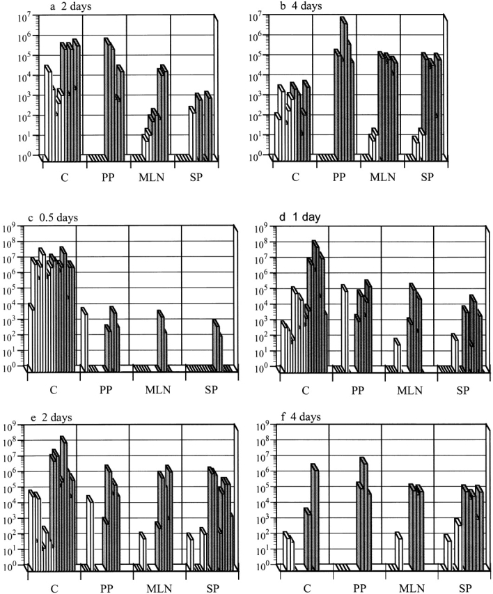
Casp-1−/− mice are resistant to S. typhimurium colonization after an oral infection. Wild-type mice (gray bars) and casp-1− / − mice (white bars) were inoculated intragastrically with wild-type S. typhimurium. The tissue colonization of mice inoculated with 2 × 10⁷ CFU of S. typhimurium SL1344 was determined for (a) 2 and (b) 4 d after inoculation. C, cecum; PP, Peyer's patches; MLN, mesenteric lymph nodes; SP, spleen. n = 5 for both days. Tissue colonization after intragastric inoculation with 1010 CFU of S. typhimurium SL1344 for (c) 0.5, (d) 1, (e) 2, and (f) 4 d after inoculation. Cecum and spleen, n = 8 for days 0.5, 1, and 2; n = 3 for day 4. PP and MLNs, n = 5 for days 0.5, 1, and 2; n = 3 for day 4. Day 1 for PP and MLNs for wild-type compared with casp-1 −/ − mice: P = 0.0236 and P = 0.0343, respectively. Day 2 for PP, MLNs, spleens, and livers: P = 0.0132, P = 0.0343, P = 0.0025, and P = .0283, respectively. Day 4 for PP, MLNs, spleens, and livers: P = 0.0369, P = 0.0038, P = 0.0207, and P = 0.0278, respectively. Rectangles on the x axis represent zero bacterial colonies present in undiluted tissue. These data represent results from one experiment, performed on age-matched casp-1 −/ − and wild-type mice, which is representative of three experiments. The variability in mouse colonization at early times is reproducible and typical of what is seen after an oral inoculation.