
Figure 3.
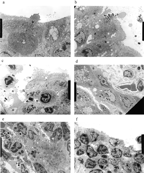
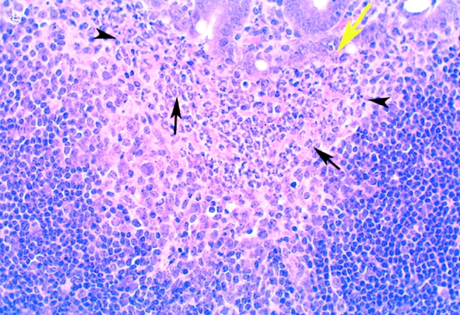
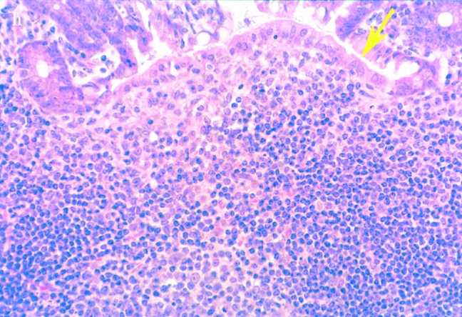
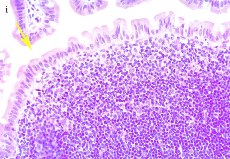
(continues on facing page). S. typhimurium induces inflammation in wild-type PP but not in casp-1 −/ − PP. Transmission electron micrographs of infected PP revealed bacterial invasion of M cells at 0.5 h in wild-type (a) and casp-1 −/ − (b) PP, and at 1 h in (c) extracellular Salmonella in wild-type and (d) intracellular bacteria in casp-1 −/ − PP. At 3 h, wild-type PP dome revealed an infiltration of PMNs and intracellular Salmonella (e), whereas casp-1 −/ − PP revealed an absence of PMNs and bacteria (f). Hematoxylin and eosin stains of PP from casp-1 +/+ (g) mice infected for 3 h revealed clusters of PMNs (black arrows) and pyknotic cells (arrowheads), whereas casp-1 −/ − PP did not (h). Uninfected, wild-type PP are shown for comparison (i). Yellow arrows point to the PP dome area. Serial tissue sections were stained with anti-Salmonella antibody to confirm that the PP shown was infected. All sections are oriented with intestinal lumen at the top and lymphoid follicle at the bottom.