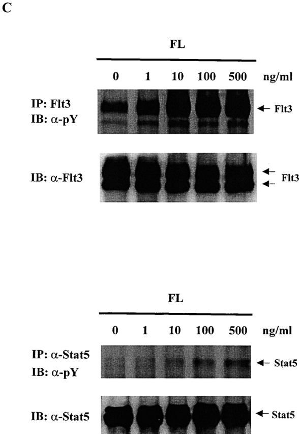
Figure 1.
FL induces tyrosine phosphorylation of Stat5 in Baf3/Flt3 cells. (A) Growth factor-starved Baf3/Flt3 cells were stimulated with FL (100 ng/ml) or IL-3 (10 ng/ml) for 5 min. Stat5 and Stat3 were immunoprecipitated from cell lysates and immunoblotted with antiphosphotyrosine antibody. (B) Stat5 is transiently tyrosine phosphorylated by FL stimulation. Growth factor–starved Baf3/Flt3 cells were stimulated with FL for various periods of time or with IL-3 for 5 min. Stat5 was immunoprecipitated from cell lysates and immunoblotted with antiphosphotyrosine antibody. (C) Dose–response of tyrosine phosphorylation of Flt3 and Stat5. Growth factor–starved Baf3/Flt3 cells were stimulated with different dose of FL for 5 min. Flt3 and Stat5 were immunoprecipitated from cell lysates and immunoblotted with antiphosphotyrosine antibody. The same membranes were stripped and reblotted with different antibodies as shown. Results shown are from one representative of two to three experiments. pY, antiphosphotyrosine antibody.