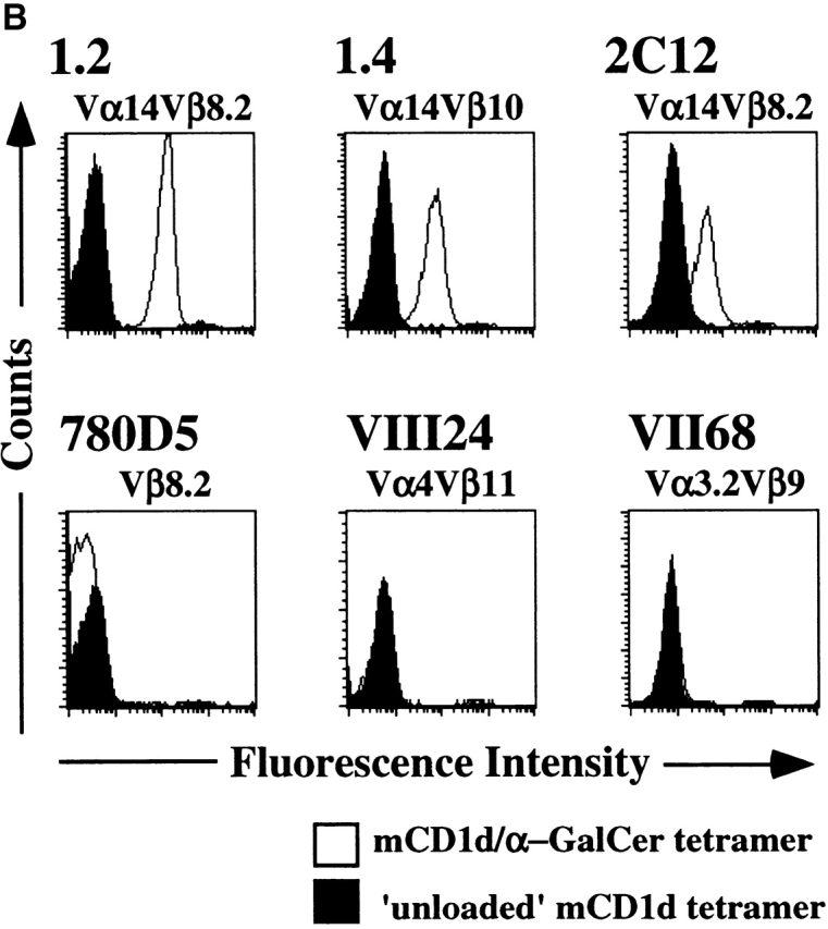
Figure 1.
Specific CD1d multimer staining of T cell hybridomas. (A) Comparison of mCD1d and hCD1d dimer staining of the 1.2 and 1.4 NK T cell hybridomas. (B) Comparison of mCD1d tetramer staining of the indicated T cell hybridomas using α-GalCer–loaded and unloaded tetramers. One representative experiment of three is shown.