
Figure 3.
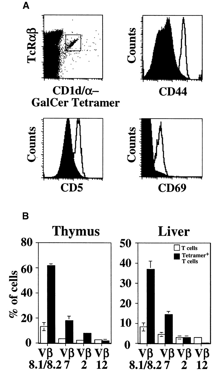
Tetramer-positive cells have the expected phenotypic markers of NK T cells. (A) Phenotypic analysis of tetramer-positive cells in the thymus. Histogram overlays of tetramer-positive TCR-βint cells (open histogram) and control stainings (shaded histogram) reveal the tetramer-positive population to be CD5+CD44highCD69+. Conventional T cells served as the control staining for CD44, and for CD5 and CD69, background staining was used due to the presence of conventional T cells staining positive for these markers. (B) Tetramer-positive T cells show a bias in Vβ gene segment usage as compared with conventional T cells. Conventional T cell controls are the lymphocytes expressing high levels of TCR-β in the thymus (upper left gate in A) and TCR-βhigh and NK1.1− T cells in the liver. Each of these figures is derived from two experiments with similar results.