Figure 4.
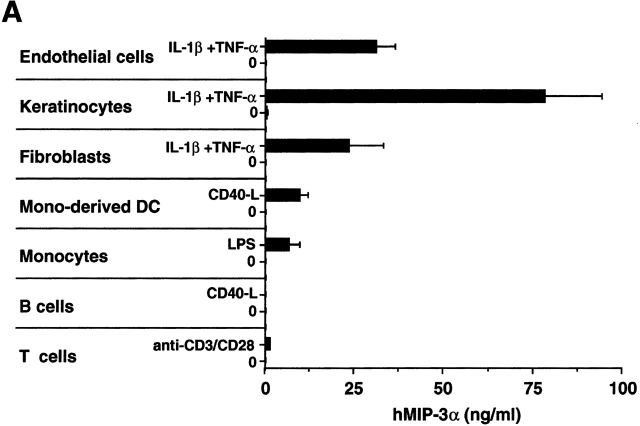
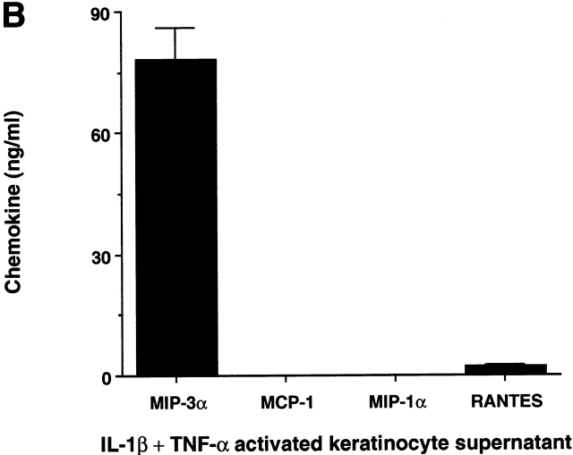
MIP-3α is selectively produced by epithelial cells. (A) Nonhematopoietic cells, including endothelial cells (human umbilical vein endothelial cells), keratinocytes, and fibroblasts, were seeded at 1–2 × 10⁴ cells/ml, and after 3–5 d of culture (80% confluence), cells were either activated by IL-1β+TNF-α or left unactivated for 48 h. Monocyte-derived DCs and B lymphocytes were either activated by CD40L-transfected L cells (murine fibroblasts, CD40L L cells) or left unactivated for 24 h. Coculture with untransfected L cells was checked for the absence of MIP-3α production. Peripheral blood monocytes and T lymphocytes were activated for 24 h in the presence of LPS and anti-CD3 plus anti-CD28, respectively. IL-1β+TNF-α did not induce MIP-3α production by monocytes or monocyte-derived DCs (not shown). Supernatants were collected and measured for MIP-3α content using a specific ELISA. (B) Keratinocyte supernatants were collected and measured for MIP-3α, MCP-1, MIP-1α, and RANTES contents using specific ELISAs. Results shown are representative of five independent experiments.


