Figure 8.
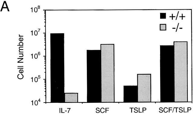
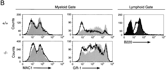
Analysis of TSLP activity in Whitlock-Witte cultures derived from wild-type or IL-7Rα−/− mice. (A) Livers from IL-7Rα−/− or wild-type newborn mice were grown under Whitlock-Witte culture conditions for 9 d in the presence of either IL-7 (10 ng/ml), SCF (1 μg/ml), TSLP (100 ng/ml), or SCF plus TSLP. Enumeration of nonadherent cells was determined by trypan blue exclusion. (B) Immunofluorescent profiles of nonadherent cells isolated from IL-7Rα−/− and wild-type mice. Lymphoid and myeloid gates were established by the forward versus side light scatter profiles of cells grown in IL-7 alone or SCF alone, respectively. Myeloid gated cells were analyzed for Mac-1 and Gr-1 expression after growth in SCF (gray histogram) or SCF plus TSLP (white histogram). Lymphoid gated cells were analyzed for B220 expression after growth in IL-7 (hatched histogram) or SCF plus TSLP (white histogram). Black histogram represents IgG1 isotype control staining of cells grown in IL-7.


