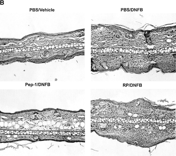
Figure 7.
Impact of Pep-1 on the expression of contact hypersensitivity responses. (A) BALB/c mice were sensitized with 0.5% DNFB on abdominal skin and challenged 7 d later with 0.2% DNFB on the right ears and vehicle alone on the left ears. These mice received the following: (a) two subcutaneous injections (40 μg/ear/injection) of the indicated peptides into both ears 24 and 1 h before challenge (n = 5); (b) a single intravenous injection (1 mg/animal) at 1 h before challenge (n = 10); or (c) a single topical application (40 μg/ear) at 1 h before challenge (n = 10). The data are representative of two independent experiments, showing the means ± SEM of the ear swelling responses at 48 h. Statistically significant differences (ANOVA for the subcutaneous injection protocol and Student's t test for other protocols) are indicated with asterisks (*P < 0.05; **P < 0.01). (B) Ear skin samples harvested from the animals receiving the subcutaneous injections of Pep-1 or RP (left columns in A) were processed for hematoxylin and eosin staining. Data shown are the representative fields (original magnification: ×100). (C) Histological samples (n = 10) were examined by a third individual for the ear thickness and the number of skin-infiltrating leukocytes under a microscope. Asterisks indicate statistically significant differences (**P < 0.01) assessed by ANOVA. (D) BALB/c mice were painted on both ears with 0.5% DNFB on day 0 and with 0.2% DNFB on days 2 and 4 (as indicated by closed triangles). Pep-1 (•) or RP (○) (40 μg/ear/injection) was injected locally in the left or right ears of the same animals, respectively, on days 0, 1, 3, and 5 (as indicated by ▿). The data shown are representative of two independent experiments, showing the means ± SEM (n = 15) of ear swelling responses. Statistically significant differences between the Pep-1 group and the RP group are indicated with asterisks (**P < 0.01; Student's t test).