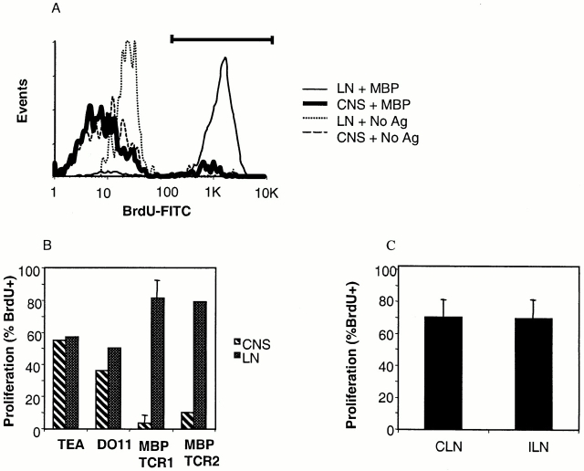
Figure 4.
CNS T cells from MBP TCR1 and MBP TCR2 transgenic mice do not proliferate in response to antigen while MBP transgenic T cells recovered from CNS-draining cervical LNs (CLN) or nondraining inguinal LNs (ILN) proliferate well. (A) Flow cytometric analysis measuring the proliferation of pooled CNS T cells purified from spinal cords of 17 MBP TCR1 transgenic mice and LN T cells from the same animals. CNS and LN T cells were separately incubated with irradiated T cell–depleted wild-type splenic APCs with and without MBP Ac 1-11. Proliferating T cells are identified as BrdU+ cells as indicated by the bar. (B) Comparison of the proliferation of CNS and LN T cells from different TCR transgenic models in response to antigenic stimulation. Experiments were performed as described in panel A and the data represent an average of three experiments using MBP TCR1 transgenic mice and one experiment each pooling 15–17 MBP TCR2, DO11.10, and TEa TCR transgenic mice. For MBP TCR1 and TEa TCR transgenic mice, proliferation of Vα2+ CNS and LN T cells is shown. For MBP TCR2 and DO11.10 TCR transgenic mice, proliferation of TCR+ rather than Vα2+ T cells is shown. (C) The percentage of proliferating (BrdU+) Vα2+ T cells was determined for T cells harvested from the CLN and ILN (n = 2). Mice used for all of these experiments were 4–12 wk of age.

