
Figure 1.
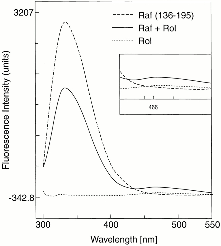
Quenching of protein fluorescence and resonance energy transfer. Fluorescence emission spectra of 250-nM solution of Gst fusion protein comprising amino acids 136–195 of human cRaf-1, excited at 280 nm in the presence (solid line) or absence (dashed line) of 250 nM all-trans retinol (Rol) and spectrum of retinol in buffer (dotted line). A fluorescence peak at 466 nm, although small, indicated energy transfer (inset). The naturally occurring trp residue at position 187 may not be optimally placed for efficient energy transfer.