
Figure 3.
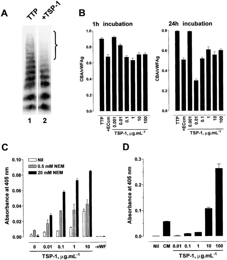
Reduction in the average multimer size of vWF by TSP-1 in vitro. (A) TTP1 patient plasma (10 μl) was incubated with Hepes-buffered saline containing 1 mM CaCl2 (TTP, lane 1) or purified platelet TSP-1 (1 μg/ml, lane 2) in the Hepes/CaCl2 buffer (90 μl) for 1 h at 37°C and aliquots of the reaction (10 μl) were resolved on 1% agarose gel electrophoresis. The vWF was transferred to PVDF membrane and Western blotted using peroxidase-conjugated anti-vWF polyclonal antibodies. The bracket highlights the change in the proportion of large vWF multimers in the population. (B) TTP1 patient plasma (10 μl) was incubated with HMEC-1–conditioned medium (+ECcm) or purified platelet TSP-1 (0.001 to 100 μg/ml) in Hepes-buffered saline containing 1 mM CaCl2 (90 μl) for 1 or 24 h at 37°C and aliquots of the reaction were analyzed for vWF antigen levels and collagen binding affinity. The results are expressed as the ratio of the collagen binding activity and vWF antigen level. The bars and errors are the mean and SD of triplicate determinations. (C) Interaction of TSP-1 with vWF. Microtiter plate wells coated with purified human vWF and blocked with BSA were incubated with purified human TSP-1 (0 to 10 μg/ml) in Hepes-buffered saline containing 1 mM CaCl2 and no (white bars), 5 mM (hatched bars), or 20 mM (black bars) NEM for 30 min at room temperature. On one occasion, wells not coated with vWF but blocked with BSA were incubated with 10 μg/ml TSP-1. The wells were washed with the Hepes buffer containing 1 M NaCl to minimize noncovalent interactions and the bound TSP-1 was meas-ured using the anti-TSP1 monoclonal antibody, HB8432, and peroxidase-conjugated secondary antibody. The bars and errors are the mean and SD of triplicate determinations. (D) Generation of new thiols in vWF by TSP-1. Purified human vWF (2 μg/ml) was incubated with Hepes-buffered saline containing 1 mM CaCl2 (Nil), HMEC-1–conditioned medium (CM), or purified human TSP-1 (0.01 to 100 μg/ml) in the Hepes/CaCl2 buffer for 60 min at 37°C. The reactions were labeled with MPB (100 μM) and the unreacted MPB was quenched with GSH (200 μM). Aliquots of the reactions were incubated in microtitre plate wells coated with anti-vWF polyclonal antibodies and the adsorbed vWF was incubated with streptavidin peroxidase to measure the incorporated MPB. The bars and errors are the mean and SD of triplicate determinations.