Figure 3.
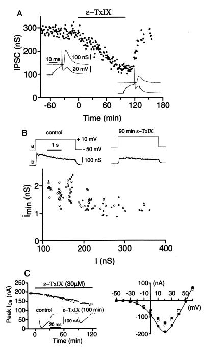
Effects of ɛ-TxIX on synaptic transmission. (A) Bath-applied ɛ-TxIX (30 μM) reduced the amplitude of IPSC. Washing out the peptide led to a rapid recovery of synaptic transmission. Insets represent the postsynaptic responses (IPSCs, Upper traces) evoked by a presynaptic action potential (Lower traces) before (Left recordings) and after (Right recordings) 100 minutes ɛ-TxIX application. (B) LDIPSCs, evoked by a 3-second depolarization of the voltage-clamped presynaptic neuron, were decreased in the presence of ɛ-TxIX (30 μM). Control response: I = 250 nS; Imin = 1.2 nS = 12.25 ms; Q = 51 020 quanta; 90 minutes ɛ-TxIX: I = 142 nS, Imin= 1.7 nS = 11.8 ms; Q = 21 236 quanta. The graph shows the relationship between the mean amplitude of MPSCs (Imin, calculated from the above LDIPSCs) and the mean amplitude (I) of the LDIPSC before (●) and after 90 minutes of ɛ-TxIX application (○). (C, Left) Evolution of the peak presynaptic Ca2+ current after bath application of ɛ-TxIX (30 μM). Insets are examples of the Ca2+ current before (Left) and after (Right) ɛ-TxIX application. (Right) I/V curves representing the presynaptic Ca2+ current in the control situation (●), after 90 minutes (○) and 115 minutes (■) of ɛ-TxIX.

