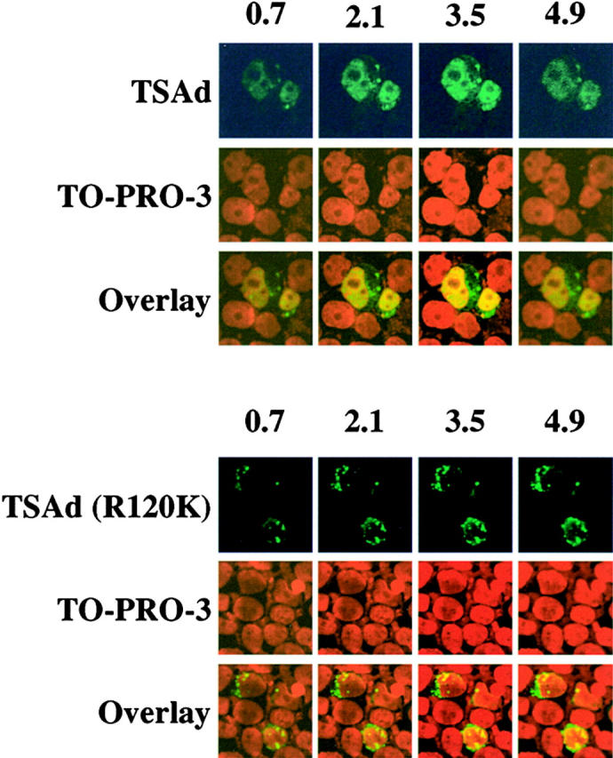
Figure 2.

Confocal microscopic analysis of TSAd nuclear expression. Expression of TSAd in 293T cells, transiently transfected with FLAG-TSAd (top) or FLAG-TSAd (R120K; bottom), was examined by immunostaining for the FLAG epitope (green) and confocal microscopy. Nuclei were revealed with the use of TO-PRO-3 (red). Serial sections, in 0.7-μm increments, from the top to bottom of samples, were viewed. Shown are select sections for each sample. The section number (in μm) is indicated at the top of columns.
