Figure 4.
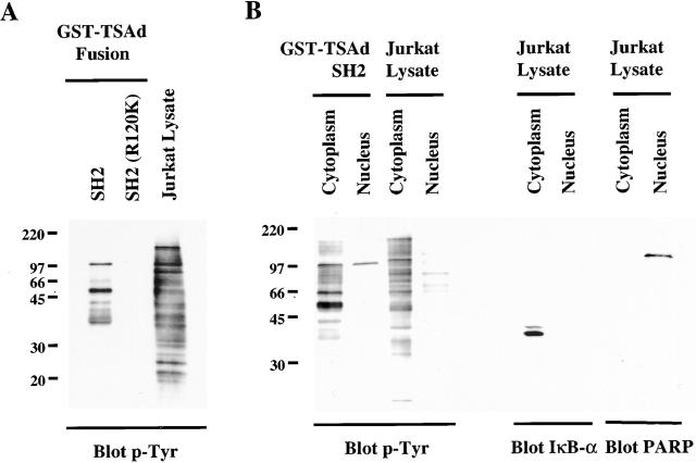
TSAd SH2 domain ligands. Glutathione agarose beads coated with GST–TSAd SH2 domain or –TSAd SH2 (R120K) fusion proteins were incubated with NP-40 lysates (A) or cytoplasmic and nuclear lysates (B) of PMA plus pervanadate-stimulated Jurkat cells (cytoplasmic and nuclear lysates were derived from the same number of cells). After extensive bead washing, fusion protein–bound tyrosine-phosphorylated proteins were eluted and detected by Western blotting using an anti-phosphotyrosine antibody (p-Tyr). In panel A, that equivalent quantities of fusion proteins were used in experiments was confirmed by Coomassie blue staining of replicate SDS-page gels (not shown). In B, blots were stripped and reprobed with anti-IκB-α and PARP antibodies (right). IκB-α was detected only in cytoplasmic lysates and PARP only in nuclear lysates indicating relative purity of the respective fractions.

