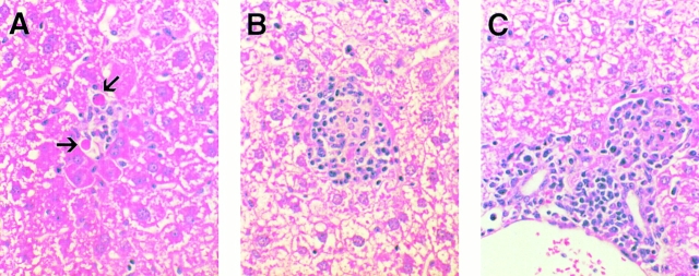
Figure 4.
Histological features of α-GalCer–induced hepatitis in the liver of mice after 10 ng of α-GalCer injection. Liver sections obtained from α-GalCer–injected mice killed on day 1 (A) or day 3 (B and C) were stained with hematoxylin and eosin as described in Materials and Methods. On day 1, the liver was mostly cytologically normal except for a few very small and widely scattered necroinflammatory foci consisting of lymphomononuclear cells and rare apoptotic hepatocytes (arrows; A). On day 3, inflammatory foci became larger and more abundant, consisting of a mixed population of lymphomononuclear cells that often displayed granulomatous features (B). In addition, a mixed lymphomononuclear cell infiltrate was also detected in and adjacent to the portal tracts at this time point (C). The infiltrate subsided progressively thereafter, returning to baseline by day 14 (not shown).

