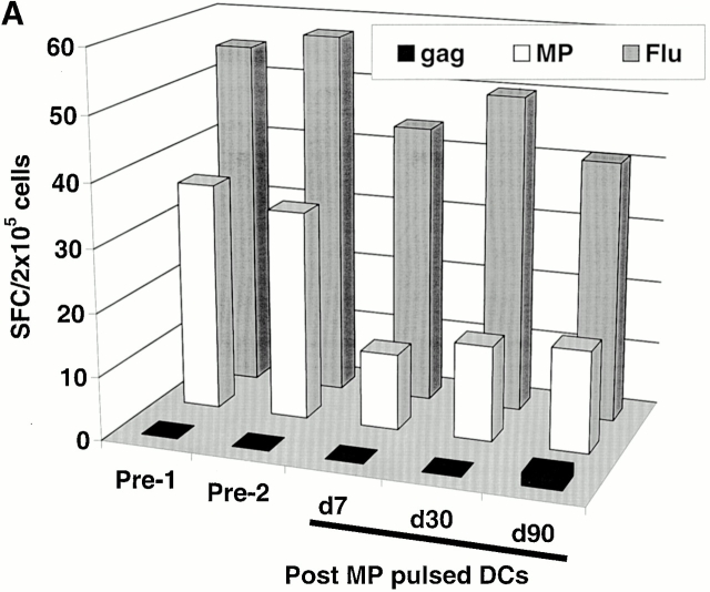
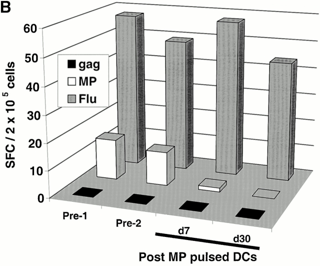
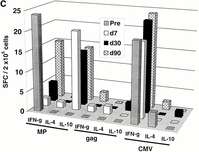
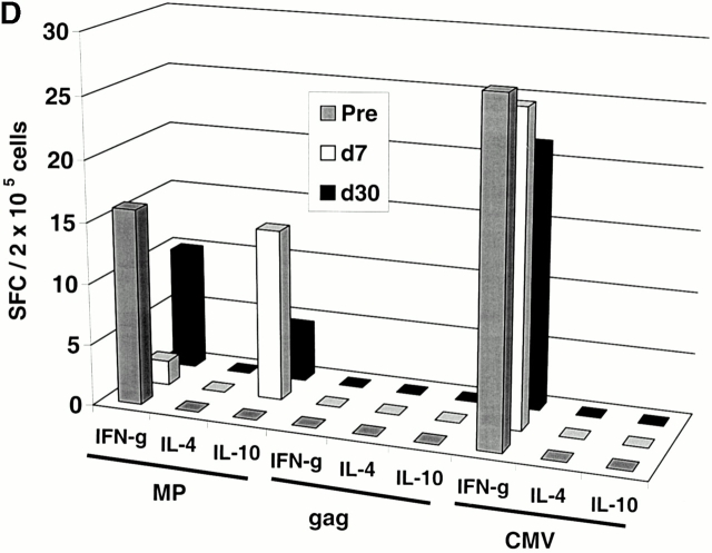
Figure 1.
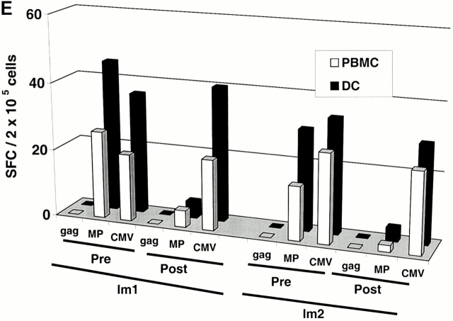
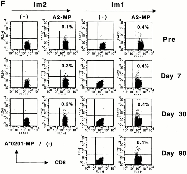
Immune responses in uncultured T cells. (A and B) MP-, gag-, and influenza (Flu)-specific IFN-γ–producing cells from before and after DC immunization were quantified in freshly isolated uncultured PBMCs using an ELISPOT assay. Data for influenza specific cells is per 105 cells. SEM for all measurements is <20%. (A) Im1; (B) Im2. (C and D). Pre- and postimmunization samples were thawed together and assayed for antigen-specific T cells secreting IFN-γ, IL-4, and IL-10 using a 16-h ELISPOT assay. Antigens were HLA A2.1–restricted peptides from influenza MP, HIV-gag (gag), and CMV pp65 (CMV). Positive controls for the assays included SEA for IFN-γ and IL-10 and PHA for IL-4 (not shown). SEM for all measurements is <20%. (C) Im1; (D) Im2. (E) Use of peptide-pulsed DCs as APCs in the ELISPOT. Pre- and postimmunization specimens were examined using peptide-pulsed mature DCs as APCs (PBMC/DC ratio 30:1) in the ELISPOT. SEM for all measurements is <20%. (F) Quantification of MP-specific T cells using MHC tetramers in uncultured cells. Pre-/postimmunization specimens were stained with A*0201–MP tetramers at 37°C and analyzed by flow cytometry. Data shown are gated for CD8+ T cells and expressed as percent CD8+ T cells binding A*0201–MP tetramer.

