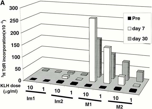
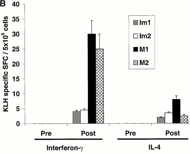
Figure 3.
Priming of KLH-specific T cells in vivo. (A) Antigen-dependent proliferation. Pre- and postimmunization PBMCs were thawed together and cultured in the absence or presence of KLH (10 μg/ml). Data shown are KLH-specific proliferation after subtracting [3H]TdR incorporation in control wells. SEM for all measurements is <30%. (B) KLH-specific IFN-γ– and IL-4–producing cells from before and after DC immunization were quantified in freshly isolated uncultured PBMCs using an ELISPOT assay. KLH-specific SFCs were calculated after subtracting data from control wells without antigen.


