
Figure 5.
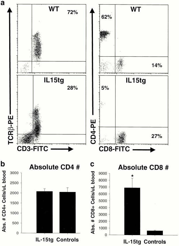
Early expansion of CD8+ T cells within IL-15tg mice. (a) Flow cytometric analysis of peripheral blood lymphocytes from representative IL-15tg and nontransgenic littermate wild-type controls (WT). Most lymphocytes in wild-type mice are CD3+TCR-β+ T cells, and the percentage of this population is reduced in IL-15tg mice due to dilution by the expanded NK cells. The CD4/CD8 ratio is significantly inverted in IL-15tg mice. (b) The absolute number of CD4+ T cells is identical in IL-15tg and control mice. (c) The absolute number of CD8+ T cells is significantly increased in IL-15tg (P < 10−4), compared with control mice. This increase in CD8+ T cells is responsible for the CD4/CD8 ratio. For b and c, data represent the mean CD4 or CD8 counts ± SEM of IL-15tg (n ± 71) and control (n = 51) mice.