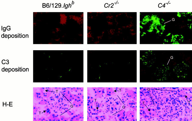
Figure 3.
Glomerular IC deposition and glomerulonephritis in C4 − /− mice. Kidney sections from 10-mo-old, female B6/129.Ighb (left), Cr2 − /− (center), and C4 − /− (right) mice were stained with FITC-conjugated goat anti–mouse IgG (green, top panels, ×200 magnification) or goat anti–mouse C3 (green, center panels, ×200 magnification) and counterstained with Evans blue (red). Glomerular cellularity was examined in sections stained with hematoxylin and eosin (bottom panels, ×400 magnification). G, glomerulus; T, tubule.

