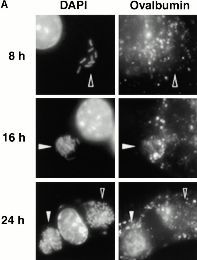
Figure 2.
The acquisition of fluid-phase lysosomal markers by L. pneumophila vacuoles. Macrophages whose lysosomes were prelabeled by endocytosis of TR-OV or TR-DEX were infected with L. pneumophila for the period indicated, fixed, incubated with DAPI to label the bacteria, then analyzed microscopically. (A) At 8 h, bacterial vacuoles were isolated from the endocytic network (black arrowheads); by 16 h, the lumen of many vacuoles harboring L. pneumophila contained a fluorescent probe (white arrowheads). The 24-h panel reveals two separate vacuoles heavily laden with bacteria; the one at left had obviously fused (white arrowheads), whereas TR-OV accumulation by the vacuole at right was below the limit of detection (black arrowheads). (B) The percentage of 50 vacuoles that contained either TR-OV (filled symbols) or TR-DEX (open symbols) at the time indicated was scored by fluorescence microscopy in four separate experiments, each represented by a different symbol (diamonds or squares).