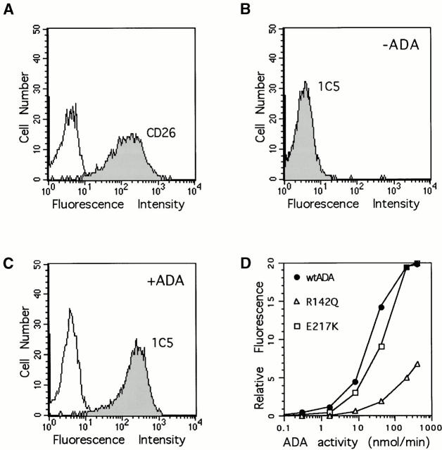
Figure 6.
Flow cytometry analysis of CD26 and ADA on the surface of AlNe cells. (A) CD26 expression. Shown are reactivity of AlNe cells with PE-conjugated anti-CD26 mAb Ta1 (shaded histogram) and control mAb IgG1-PE (open histogram). (B and C) ADA. Anti-ADA mAb 1C5 binding to AlNe cells that had been washed after incubation with lysate of untransformed SØ3834 (B) or with lysate of SØ3834 expressing wild-type human ADA (400 nmol/min per ml of medium) (C). (D) Binding of wild-type (wt) human ADA (circles), R142Q (triangles), and E217K (squares) ADA mutants. AlNe cells were incubated with 0.3, 1.7, 8.3, 42, 209, and 400 nmol/min per ml of recombinant wild-type and R142Q ADAs. The amounts of E217K-expressing SØ3834 lysate protein used (0.024–30 μg) were made equal to wild-type ADA lysate protein. After washing, cell surface–associated ADA was determined by reactivity of mAb 1C5 and flow cytometry. For clarity, the horizontal axis shows only units of ADA activity. Data shown are from one of two experiments.

