
Figure 3.
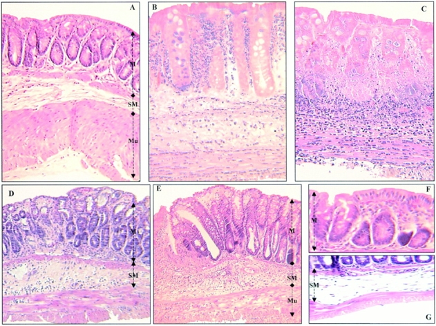
Representative histological sections of colon tissues of Balb/c mice. (A) Normal transparietal colon section of a vehicle-treated mouse with an Ameho score of 0 (×250). The different layers are indicated: M, mucosa; SM, submucosa; Mu, muscular layer. (B) Transparietal colon section (Ameho score 6) 2 d after the induction of colitis by TNBS. Thickening of the colon wall, with a predominant inflammatory infiltrate in the lamina propria, and necrosis extending deeply into the muscular and serosal layers are evident (×400). (C) Transparietal colon section (Ameho score 6) 5 d after the induction of colitis by TNBS. Parietal necrosis extending deeply into the muscular layer with the disappearance of cells in the mucosa is visible (×250). (D) Transparietal colon section of a mouse, which received rosiglitazone before TNBS administration. The mouse was killed 2 d after colitis induction. The Ameho score was graded 2. The picture shows a subepithelial edema with a diastasis of the crypts and a moderate inflammatory infiltrate in the mucosa and submucosa (×250). (E) Transparietal colonic section of mice treated with rosiglitazone after administration of TNBS. The pictures show mice killed 5 d after colitis induction. Ulceration extending into the submucosa, associated with a mucosal, submucosal, and muscular inflammatory infiltrate involving <50% of the specimen, is visible (×250). (F and G) Colon sections of mice that received rosiglitazone before TNBS administration. The mice were killed 2 d after colitis induction. In some cases, a total repair of the mucosa was observed (F; ×600) despite the persistence of an in-depth focal necrosis in the submucosal layer (G; ×400).