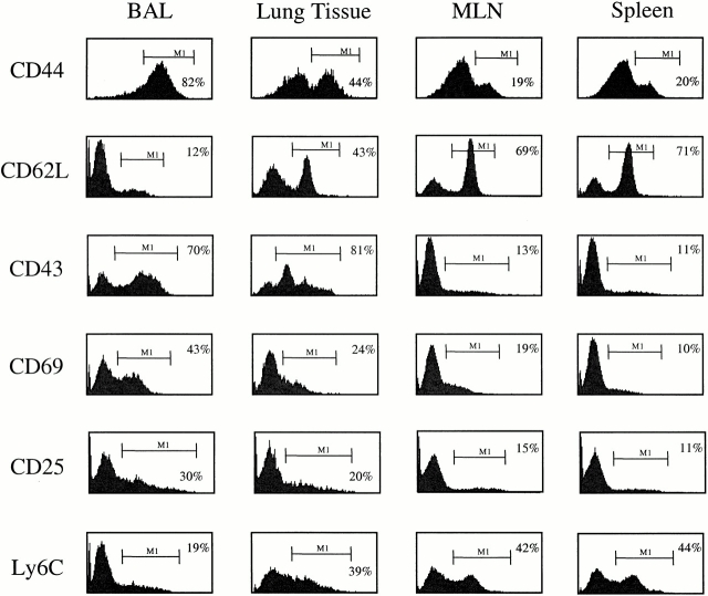
Figure 1.
CD4+ T cells in the lung airways (BAL) and lung tissues express an activated phenotype. Cells were isolated from the spleen, MLNs, lung tissue, and lung airways (BAL) of mice that had recovered from a Sendai virus infection (day 41 after infection). After isolation, the cells were stained with anti-CD4–PE and FITC-conjugated CD44, CD62L, CD43, CD69, and Ly6C antibodies. Cell surface expression of CD25 was determined using anti-CD25–PE and anti-CD4–FITC antibodies. The histograms show the expression of the indicated markers among live CD4+ lymphocytes. The bars and numbers in each panel show the percentage of CD4+ cells expres-sing the indicated marker. Data are representative of four independent experiments.

