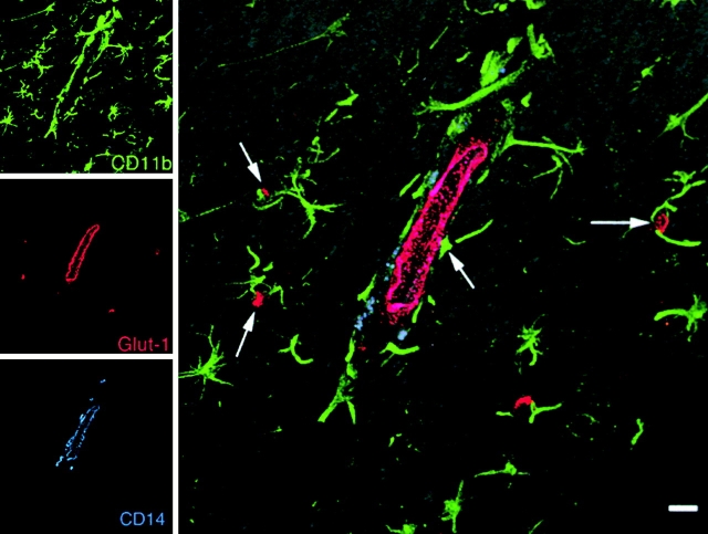
Figure 5.
Triple-label confocal microscopy of the brain of an SIV-infected macaque. Images for individual channels (CD11b, green; Glut-1, red; and CD14, blue) are shown on the left and a larger merged image contain all three channels plus the differential interference contrast (DIC) image are shown on the right. Parenchymal microglia (CD11b, green) maintain a reticular network in the white matter of SIV-infected macaques. Perivascular macrophages (CD11b+CD14+, blue-green) are situated around CNS endothelium (Glut-1, red). Both parenchymal microglia (green) and perivascular macrophages (blue-green) are in contact with CNS endothelium where parenchymal microglia have foot processes on endothelium (arrows) and perivascular macrophages are in contact with and wrap around CNS vessels. Bar, 10 μM.

