
Figure 7.
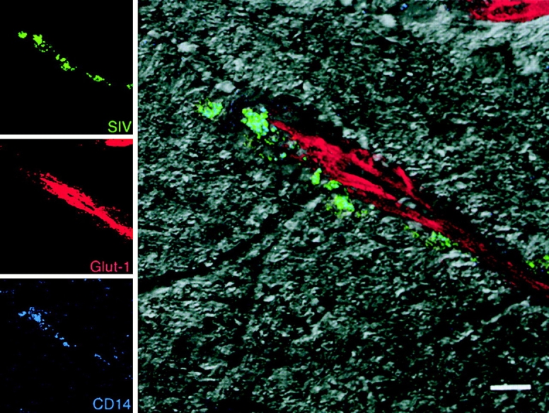
SIV-infected perivascular macrophages. Images for individual channels (SIV-p28, green; Glut-1, red; and CD14, blue) are shown on the left, and the merged image combining all three channels plus the DIC image is shown on the right. Perivascular macrophages (CD14, blue) that are viral protein positive (SIV-p28, green) appear blue-green near a CNS vessel (Glut-1, red). Bar, 10 μM.