
Figure 2.
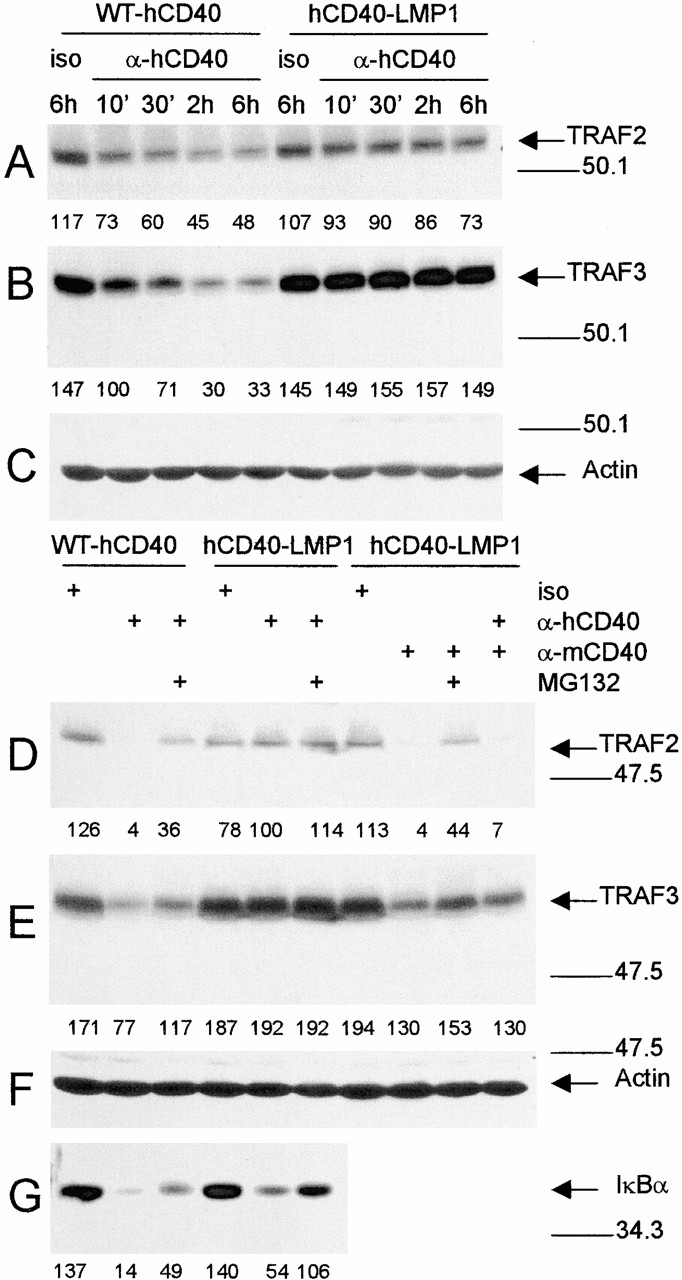
TRAF degradation after ligation of WT-hCD40 or hCD40-LMP1. (A–C) Expression-matched M12.4.1 subclones stably transfected with WT-hCD40 or hCD40-LMP1 were stimulated for the indicated number of minutes or hours with anti-hCD40 or isotype control mAbs (iso), as described in Materials and Methods. Total cell lysates were prepared and analyzed as in the legend to Fig. 1, using blotting Abs specific for (A) TRAF2, (B) TRAF3, or (C) actin. (D–G) Stable transfectants of M12.4.1 cells expressing matched amounts of the molecules indicated at the top of the lanes were stimulated for 2 h (D–F) or 20 min (G) with either anti-hCD40, anti-mCD40, or isotype control mAbs appropriate for each, as indicated to the left at the top of the figure. Where indicated, stimulation was performed in the presence of the proteasome inhibitor MG132. Total cell lysates were prepared and analyzed as above. Blotting Abs were specific for (D) TRAF2, (E) TRAF3, (F) actin, and (G) IκBα. Densitometric values are indicated below each band. Data presented are representative of three independent experiments performed with two sets of stably transfected subclones.