
Figure 4.

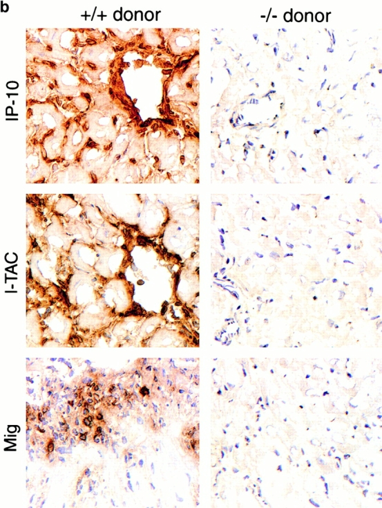

Mechanisms underlying the beneficial effects of targeting donor tissue IP-10 expression. (a) Histology showing acute rejection with extensive mononuclear cell infiltration, vascular injury, and myocyte necrosis in grafts from IP-10+/+ donors versus almost normal histology in grafts from IP-10−/− donors (day 7 after transplant). Paraffin sections, hematoxylin; original magnifications: ×300. (b) Immunohistologic detection of IP-10, I-TAC, and Mig in cardiac allografts from IP-10+/+ but not IP-10−/− donors (day 7 after transplant). Cryostat sections, hematoxylin; original magnifications: ×450. (c) Immunohistologic analysis showed significant reduction in recruitment of intragraft CD45+ cells (all leukocytes), CD4 and CD8 T cell subsets, macrophages, and IL-2R+ (CD25+) cells in allografts from IP-10−/− versus IP-10+/+ donors at day 7 after transplantation. Data (mean ± SD) from counting of 20 consecutive fields/graft and 3 grafts/group. *Significantly reduced cell numbers versus controls (*P < 0.05; **P < 0.01; ***P < 0.005).