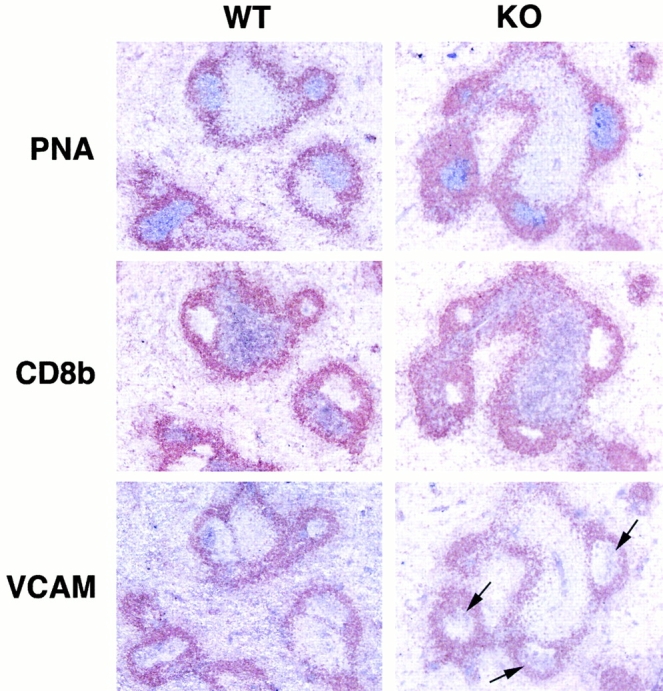
Figure 6.

Relatively normal B cell responses in vcam-1flox/Δ/TIE2Cre+ mice. Serial spleen sections from a vcam-1flox/flox mouse (WT) and a vcam-1flox/Δ/TIE2Cre+ mouse (KO) 10 d after intraperitoneal challenge with NP13CG adsorbed to alum (original magnifications: ×100). All sections were stained with anti-IgD antibody (seen in brown) and either PNA, anti-CD8b, or anti–VCAM-1. PNA, CD8b, and VCAM-1 staining are in purple. Arrows in the KO section indicate typical VCAM-1 staining on FDCs within GCs.
