
Figure 1.
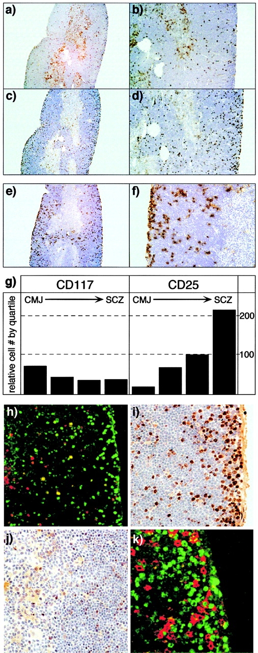
Anatomic localization of early precursors in the adult thymus. Low magnification (a and c, original magnifications: 40×) and high magnification (b and d, original magnifications: 100×) views of serial sections of adult thymus, stained with anti-CD117 (brown, a and b) or anti-CD25 (c and d) antibodies. The counterstain (blue) is hematoxylin. CD117+ cells are most frequent near the CMJ and are also scattered at lower density throughout the cortex, but are relatively infrequent in the SCZ. In contrast, CD25+ cells are rare in the vicinity of the CMJ, but increase in frequency with proximity to the SCZ. The total anatomic range for CD4−8− precursors is seen in e (original magnification: 40×) and f (original magnification: 200×), using a congenic marker for RAG-2–deficient cells (Thy-1.2) in thymuses from RAG-2/wild-type (Thy-1.1+) bone marrow chimeras. The relative distribution of CD117+ or CD25+ cells in the cortex, divided into quartiles, is illustrated in g; data are derived from counting multiple CD25/117 serial sections. h shows two-color immunofluorescent staining of CD117 (red) and CD25 (green) in a single cortical field (original magnification: 200×; left, CMJ). DN2 cells (yellow, CD117+CD25+) are generally found in the midcortical region. i and j illustrate the location of proliferating cells (brown, anti-BrdU) in the SCZ/outer cortex or inner cortex/CMJ, respectively (original magnification: 400×) after a single 4-h pulse of BrdU. As reported previously, the vast majority of cell proliferation is found to occur in the SCZ, although infrequent BrdU+ cells are found throughout the cortex. However, two-color staining (k) with CD25 (red) and BrdU (green) reveals that most of the proliferating cells in the SCZ are not DN cells (CD25+), and therefore represent amplification of the early DP compartment (preDP).