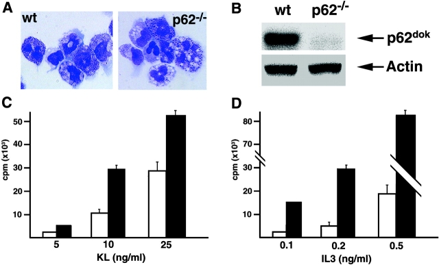
Figure 2.
Effect of p62dok disruption on BMMCs proliferation. (A) Morphology of wild-type (wt) and p62dok−/− BMMCs. (B) Expression of p62dok and actin detected by Western blot analysis in protein extracts (60 μg) from BMMCs. (C and D) Proliferative response, determined as [3H]thymidine incorporation, of wild-type and p62dok−/− BMMCs upon KL and IL-3 stimulation. Wild-type, white bar; p62dok−/−, black bar. All graphs are representative of experiments repeated three to five times. Error bars represent standard deviation.

