
Figure 1.


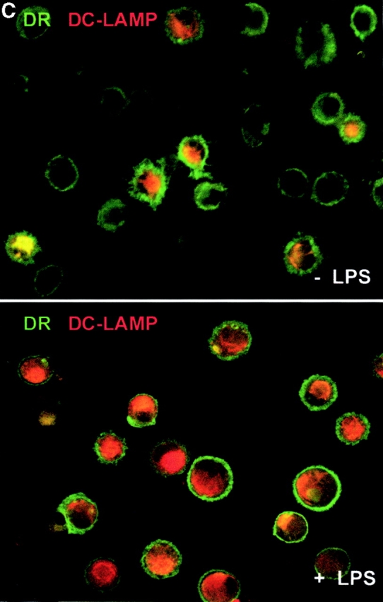


IL-15 and GM-CSF skew monocyte differentiation into DCs with features of LCs. (A) Purified monocytes are cultured with GM-CSF/IL-4 (IL4-DCs) or with GM-CSF/IL-15 (IL15-DCs) for 6 d. Both cultures display similar SSC/FSC properties (left panels). IL15-DCs acquire CD1a, lose CD14, and are HLA-DR+ (right panels), similarly to IL4-DCs (not shown). (B) IL15-DCs (bold line) acquire a mature DC phenotype (CD40high, CD83+, CD80high, CD86high, and HLA-DRhigh), when cultured with LPS (100 ng/ml) (dotted line). Thin solid line shows isotype control. (C) Some IL15-DCs express intracellular DC-LAMP, the expression of which is considerably upregulated by LPS-activation (confocal microscopy, field 130 × 100 μm). (D) IL15-DCs, but not IL4-DCs, express the surface phenotype of LCs expressing E-Cadherin, CCR6, and Langerin. Conversely, IL4-DCs, but not IL15-DCs, express high levels of CD9, CD11b, CD32, and CD36. Purified monocytes are cultured for 6 d, stained with indicated Abs and analyzed by flow cytometry. (E) Immunoblot-detection of Langerin in IL15-DCs. Proteins of SDS-gels were transferred onto Immun-Blot® PVDF membranes and these were stained using anti-Langerin and ECL Western blotting detection system. Lane 1: T cells; lane 2: IL4-DCs; lane 3: IL15-DCs. (F) Monocytes cultured with GM-CSF/IL-15 display high Langerin expression when compared with monocytes cultured with GM-CSF/IL-4/TGF-β1 (representative of four experiments).