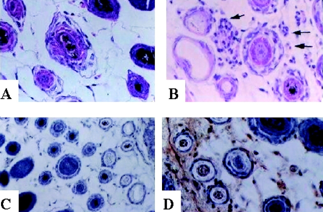
Figure 1.
Histological signs of autoimmunity in depigmented skin of mice surviving B16 challenge. Transverse sections from depigmented (B and D) or uninvolved skin (A and C) from the same mouse were stained with hematoxylin and eosin to detect cellular infiltrate (A and B), or stained for the presence of Ig deposition (C and D) following procedures described in Materials and Methods.

