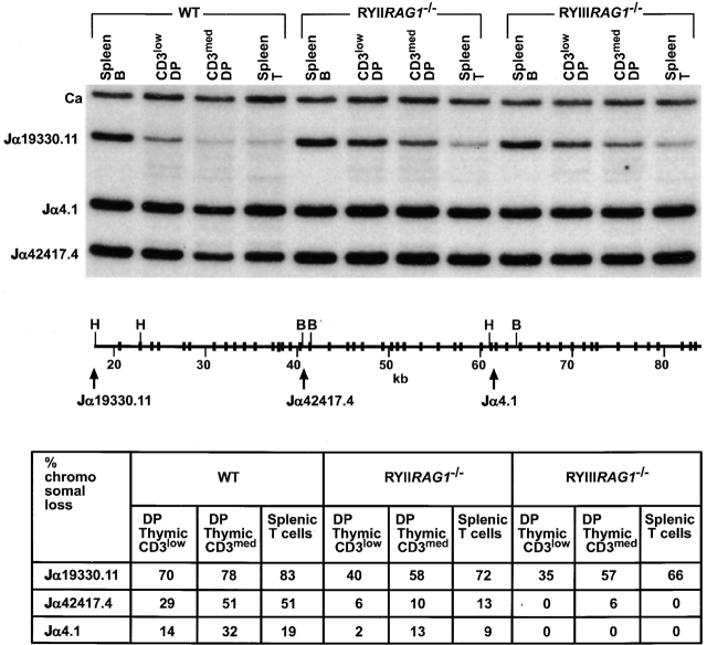
Figure 5.
Quantitation of V-Jα rearrangement by Southern blotting. CD4+ CD8+CD3low and CD3med populations were purified from thymus by flow cytometry; splenic T and B cells were purified from spleen. DNA was digested with BamHI (B) and HindIII (H). The probes are 5′ (Jα19330.11), middle (Jα42417.4), 3′ (Jα4.1), and Cα (reference 28). Schematic representation of the Jα locus showing only the relevant restriction sites; the position of the Jα probes is indicated with arrows. Jα hybridization to spleen B cell DNA was used to calculate the relative loss of Jα DNA in purified T cells. The results in the table are from one of two experiments, the variation between experiments was <5%.

